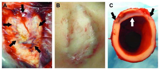
Figure 3.

Thoracic aorta patched with the biomaterial after 6 months of implantation. a) The patch still in vivo. The suture material can still be seen on the adventitial layer of the vessel. There is minimal scar tissue such that the patch is visible through it. The arrows indicate the outline of the patch; b) The lumen of the aorta with the patch. There are no blood clots associated with the patch and grossly it has integrated smoothly with the host tissue; c) A cross sectional view of the aorta through the patch showing the smooth transition between patch and native tissue.
