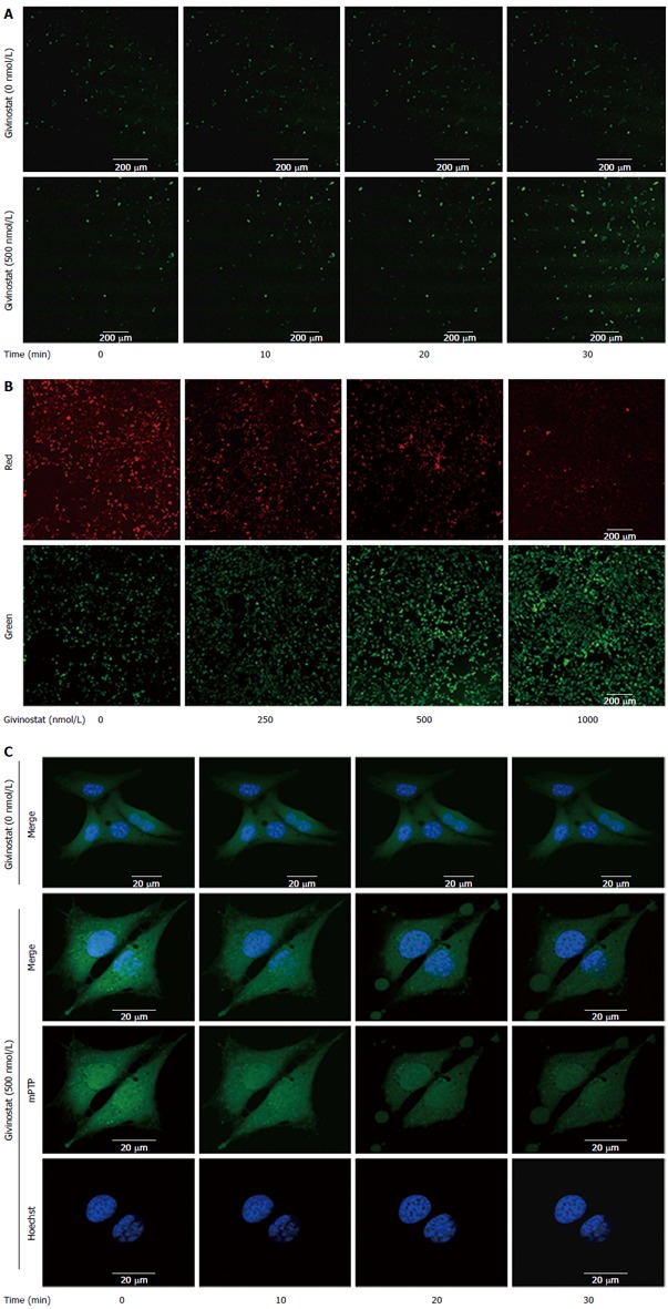
Figure 3.

Effects of givinostat on the reactive oxygen species profile, mitochondrial membrane potential, and mitochondrial permeability transition pore opening in JS-1 cells. A: Effects of givinostat on reactive oxygen species (ROS) in JS-1 cells were determined by 2’,7’-dichlorofluorescein diacetate. Green fluorescence represents the intracellular ROS. Givinostat significantly increased ROS production, in particular at 30 min (P < 0.01); B: Mitochondrial membrane potential was detected using the fluorescent probe JC-1. The green and red fluorescence represented the low and high membrane potentials, respectively. Givinostat reduced mitochondrial membrane potential in a concentration-dependent manner, particularly at 1000 nmol/L (P < 0.01); C: The mitochondrial permeability transition pore (mPTP) opening was detected by coloading with calcein-AM and CoCl2. In the givinostat treatment group, considerable mPTP opening was observed, along with decreased intensity of fluorescence in comparison to that of the control group.
