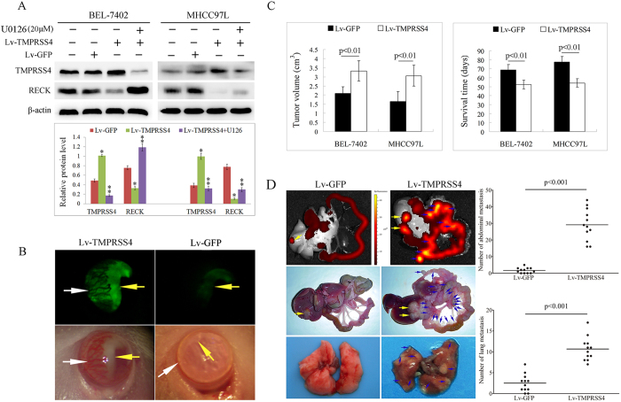
Figure 3.
A Western blot analysis revealed that cells transfected with TMPRSS4 showed significantly reduced expression of RECK, and ERK1/2 inhibitor of U0126 reversed the effect of TMPRSS4 on RECK expression; B TMPRSS4 overexpression HCC cells formed much bigger tumors in the anterior chamber of mice (yellow arrow) with large amounts of angiogenesis induced (white arrow); CThe tumors of the Lv-TMPRSS4 group were significantly bigger than those of the Lv-GFP group (p < 0.01), and the survival time of Lv-TMPRSS4 group was significantly longer than that of Lv-GFP group (p < 0.01); D The mice bearing Lv-GFP tumors showed small luminescence in the liver (yellow arrow) and no luminescence in the peritoneum, whereas mice bearing Lv-TMPRSS4 tumors demonstrated strong luminescence both in the liver (yellow arrow) and in the peritoneum (blue arrow); Pathological examination also revealed that both abdominal and lung metastasis (blue arrow) were significantly higher in Lv-TMPRSS4 group than in Lv-GFP group (p < 0.001). Bars, SD.

