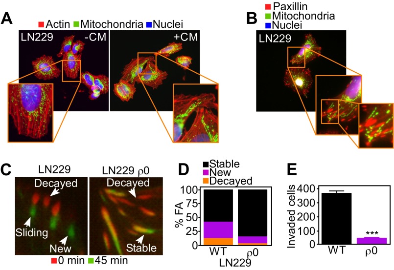
Fig. S7.
Requirement of mitochondrial respiration for FA turnover. (A and B) Respiration-competent (WT) LN229 cells were incubated with Mitotracker Red, phalloidin Alexa488, and DAPI (A) or labeled for FA complexes with an antibody to paxillin (B) and analyzed by fluorescence microscopy. Magnification, 60×. CM, NIH3T3 conditioned media. (C) WT LN229 or mtDNA-depleted LN229 (ρ0) cells expressing Talin-GFP to label FA complexes were analyzed by time-lapse microscopy. Representative images at initial (0 min) and final (45 min) time points of the analysis were merged to visualize FA turnover. Representative images of the different stages of FA complexes (decayed, sliding, stable, or new) are indicated. (D) WT or mtDNA-depleted LN229 (ρ0) cells expressing Talin-GFP were analyzed by time-lapse microscopy and quantitated for decay, formation, and stability of FA complexes per cell over 45 min; n ≥ 431. See also Movie S3. (E) WT or ρ0 LN229 cells were analyzed for invasion across Matrigel-coated Transwell inserts. Mean ± SEM (n = 2). ***P < 0.0001.

