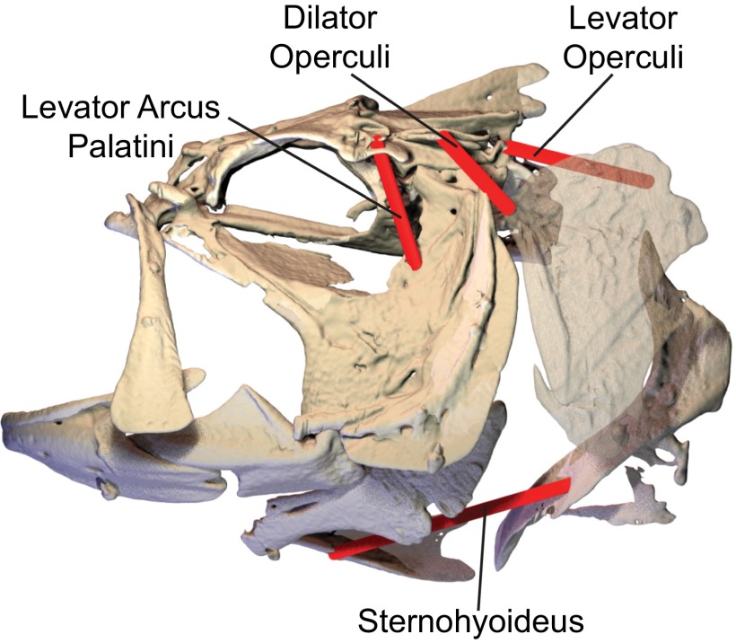
Fig. S1.
Measurement of cranial muscle lengths, using X-ray reconstruction of moving morphology (XROMM) animations. For each of the four cranial muscles shown above, the length of a representative fiber (red cylinder) was measured from XROMM animations by calculating the distance between the bony attachment sites of each fiber.

