Fig. 2.
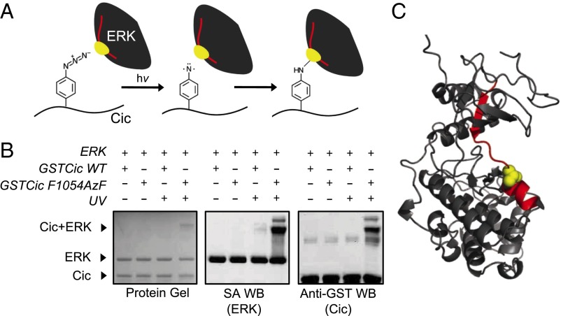
Photocrosslinking of ERK and AzF-incorporated Drosophila Cic and identification of crosslinking site by mass spectrometry. (A) Schematic representation of Cic–ERK photocrosslinking chemistry. The ERK tryptic peptide and amino acid that are crosslinked to Cic are represented in red and yellow, respectively. (B) GSTCic WT and F1054AzF were incubated with biotinylated ERK and exposed to UV light. The formation of a slow migrating, crosslinked ERK–GSTCic heterodimer was determined by protein gel, SA Western blot (detects biotinylated ERK), and anti-GST Western blot (detects GSTCic). (C) Location of crosslink on ERK. The ERK tryptic peptide found crosslinked to the Cic AzF-containing peptide is shown in red on the ERK crystal structure. The Cic-linked residue (T108) is shown in yellow in space-filling representation. Structure is drawn from Protein Data Bank (PDB) file 1ERK (27).

