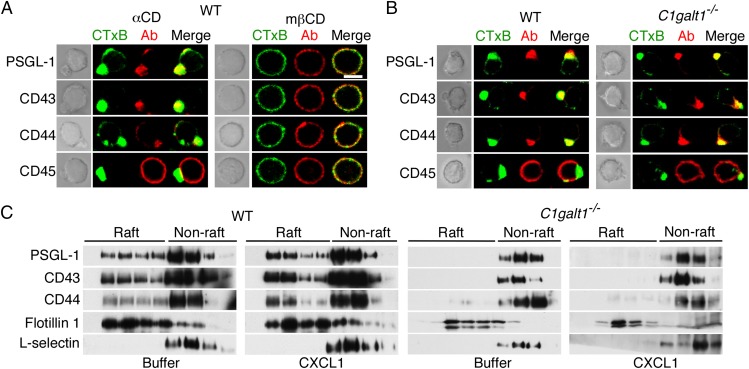
Fig. 5.
PSGL-1, CD43, and CD44 do not require core 1-derived O-glycans to redistribute to the uropods of polarized neutrophils. (A) WT neutrophils preincubated with methyl-β-cyclodextrin (mβCD) or control α-cyclodextrin (αCD) were stimulated with the chemokine CXCL1. After fixation, the cells were labeled with CTxB (green) or antibodies to the indicated protein (red). Representative cells were visualized by phase-contrast microscopy (Left) or confocal microscopy (Right). (B) WT or C1galt1−/− neutrophils were stimulated with CXCL1, fixed, labeled, and visualized as in A. (C) WT or C1galt1−/− neutrophils were incubated with buffer or CXCL1. The cells were lysed, fractionated on OptiPrep gradients, and analyzed by Western blotting with antibodies to the indicated protein as in Fig. 1. Results are representative of at least three experiments. (Scale bar, 5 μm.)

