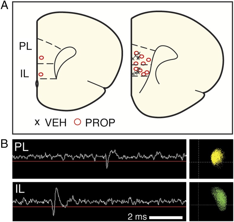
Fig. 1.
In vivo mPFC recordings in freely moving rats. (A) Histological localization of the center of each electrode array in the mPFC; each array targeted both PL (eight wires) and IL (eight wires). Right hemisphere, coronal sections represent (left to right) coordinates +3.2 and +2.7 relative to bregma in the anteroposterior plane. Six rats received propranolol (PROP) treatment, and five received vehicle (VEH) treatment. In one of the propranolol rats, recordings were obtained only from PL. (B) Example voltage trace from an electrode in PL (Upper) and IL (Lower), showing an action potential and its corresponding principal component scatter plot.

