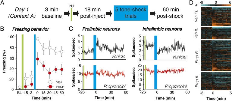
Fig. 2.
Propranolol stabilizes single-unit firing in mPFC neurons after footshock stress. (A) Day 1 experimental design. (B) Propranolol-treated rats (red circles) exhibited reduced freezing throughout the day 1 recording session relative to vehicle-treated controls (white circles). (C) Four representative histograms (20-s bins) showing spontaneous firing rate from neurons recorded in PL (Left) and IL (Right). Fear conditioning (blue bar) altered the firing rate of the PL and IL neurons obtained from vehicle-treated rats (black traces, Upper) and propranolol administration mitigated this effect (red traces, Lower). (D) Normalized firing rate heat maps showing postconditioning increases (light orange) and decreases (light blue) relative to baseline (preconditioning) firing rate (black) for all of the units recorded in each group and brain region. Only the 3 min before conditioning and the first 5 min after conditioning are shown for clarity. In both PL and IL, single units in vehicle-treated rats exhibited increases or decreases in firing rate after conditioning, and propranolol treatment mitigated these effects. Injection (INJ) is denoted by green vertical bar; conditioning (tone-shock pairings) is denoted by blue vertical bar. Data during the conditioning period were not recorded. All values are means ± SEM for freezing.

