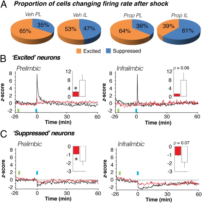
Fig. 4.
Propranolol stabilizes both increases and decreases in mPFC firing rate. (A) Proportion of neurons in each of the four groups exhibiting immediate postconditioning (first 20-s bin) increases (z > 0, excited) or decreases (z < 0, suppressed) in spontaneous firing rate. Regardless of drug treatment, PL neurons tended to increase their firing rate after shock more so than IL neurons. (B and C) Normalized firing rate histograms for PL (Left) and IL (Right) neurons that were excited (B) or suppressed (C) in the immediate postshock period in vehicle- (black traces) and propranolol-treated (red traces) rats. Inset graphs show the values for the first 20-s postshock bin in vehicle- (white bars) and propranolol-treated (red bars) rats. Injection (INJ) is denoted by green vertical bar; conditioning (tone-shock pairings) is denoted by blue vertical bar. Data during the conditioning period were not recorded. *P < 0.05 vs. vehicle. All values are means (±SEM in insets).

