Fig. 2.
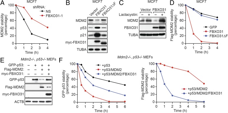
FBXO31 directs degradation of MDM2. (A) Quantification of a cycloheximide-chase/immunoblot assay monitoring MDM2 stability in MCF7 cells expressing NS or FBXO31 shRNA following treatment with cycloheximide. The graph shows the ratio of the relative levels of MDM2 and PCNA (control) at each time point; time 0 was set to 100%. (B) Immunoblot monitoring MDM2, p53, and p21 in MCF7 cells expressing empty vector, FBXO31, or FBXO31∆F. (C) Immunoblot monitoring MDM2 in MCF7 cells expressing vector or FBXO31 and treated in the presence or absence of lactacystin. (D) Quantification of a cycloheximide-chase/immunoblot assay monitoring Flag-MDM2 stability in cycloheximide-treated MCF7 cells expressing GFP (control), FBXO31, or FBXO31∆F. (E) Immunoblot monitoring GFP-p53, Flag-MDM2, and myc-FBXO31 in Mdm2−/−, p53−/− MEFs coexpressing combinations of p53, MDM2, and FBXO31. β-Actin (ACTB) was monitored as a loading control. (F) Quantification of a cycloheximide-chase/immunoblot assay monitoring GFP-p53 and Flag-MDM2 stability in cycloheximide-treated Mdm2−/−, p53−/− MEFs coexpressing combinations of p53, MDM2, and FBXO31.

