Fig. S1.
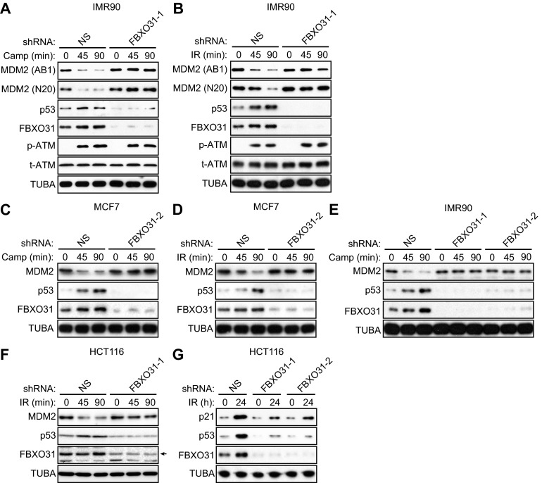
Confirmation of the results in Fig. 1 in other p53-positive cell lines and using a second FBXO31 shRNA. (A and B) Immunoblot analysis monitoring levels of MDM2 [using a monoclonal (AB1) or polyclonal (N20) antibody], p53, FBXO31, phosphorylated ATM [p-ATM(1981)], and total ATM (t-ATM) in IMR90 cells expressing NS or FBXO31 shRNA and treated in the presence or absence of camptothecin (A) or γ-irradiation (B). α-Tubulin (TUBA) was monitored as loading control. (C and D) Immunoblot analysis monitoring levels of MDM2, p53, and FBXO31 in MCF7 cells expressing NS or FBXO31 shRNA (unrelated to that used in Fig. 1 A and B) and treated in the presence (45 or 90 min) or absence (0 min) of camptothecin (C) or γ-irradiation (D). (E) Immunoblot analysis monitoring levels of MDM2, p53, and FBXO31 in IMR90 cells expressing NS or FBXO31 shRNA (unrelated to that used in A) and treated in the presence or absence of camptothecin. (F) Immunoblot analysis monitoring levels of MDM2, p53, and FBXO31 in HCT116 cells expressing NS or FBXO31 shRNA and treated in the presence or absence of γ-irradiation. (G) Immunoblot analysis monitoring levels of p21, p53, and FBXO31 in HCT116 cells expressing NS or one of two unrelated FBXO31 shRNAs and treated in the presence or absence of γ-irradiation.

