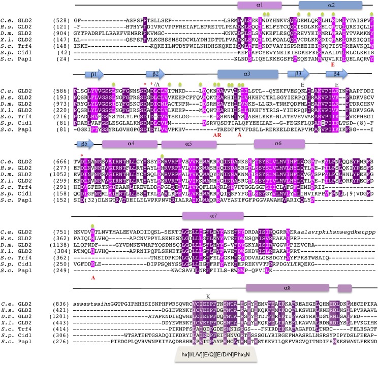
Fig. S3.
Structure-based sequence alignment of GLD-2 orthologs from C. elegans (C.e.), Homo sapiens (H.s.), Drosophila melanogaster (D.m.) and Xenopus laevis (X.l.) together with S. cerevisiae (S.c.) Trf4, S. pombe (S.p.) Cid1, and S.c. Pap1. The secondary structure elements of C.e. GLD-2 are shown above the sequence and colored according to Fig. 1A. Strictly and well conserved residues are highlighted in dark and light purple, respectively. GLD-3–interacting residues are indicated with green dots. Residues targeted for mutagenesis are shown with the substituted amino acid in red, below the sequences, or black, above the sequences. Active-site residues discussed in the text are indicated with an asterisk. Also indicated is the sequence corresponding to the nucleotide recognition motif (NRM; refs. 28 and 40).

