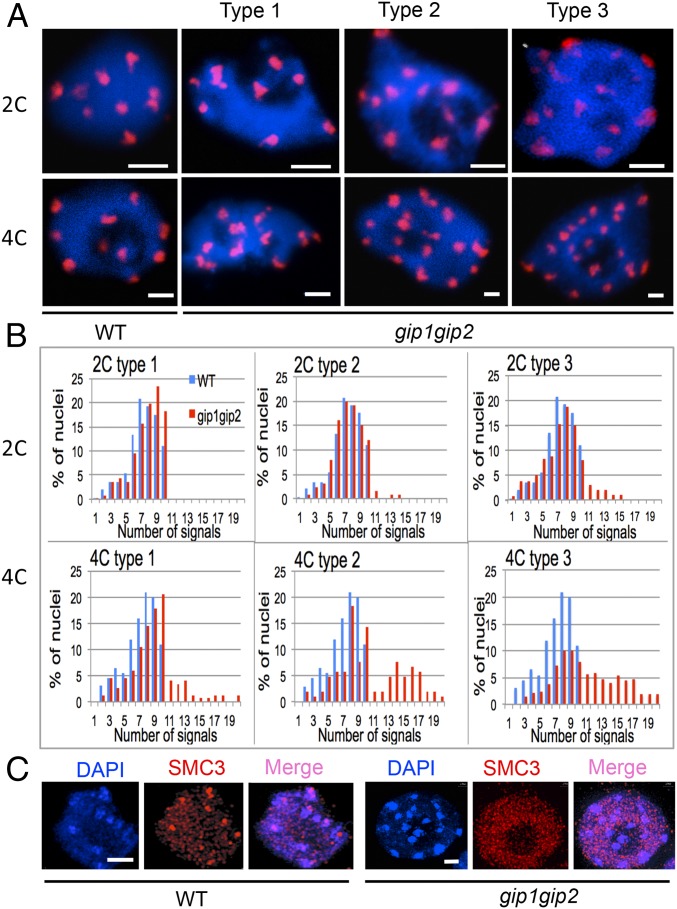
Fig. 1.
gip1gip2 mutants exhibit centromeric cohesion defects. (A) FISH detection of centromeric pAL signals in 2C and 4C flow-sorted nuclei from WT and three seedling phenotypes (types 1–3) of gip1gip2 mutants. (B) Number of pAL signals in nuclei (2C and 4C WT, n = 120; gip1gip2 type 1, n = 116; type 2, n = 122; type 3, n = 125). (C) Immunolocalization of the SMC3 cohesin subunit in meristematic root nuclei. DAPI staining is shown in blue. (Scale bars, 2 µm.)

