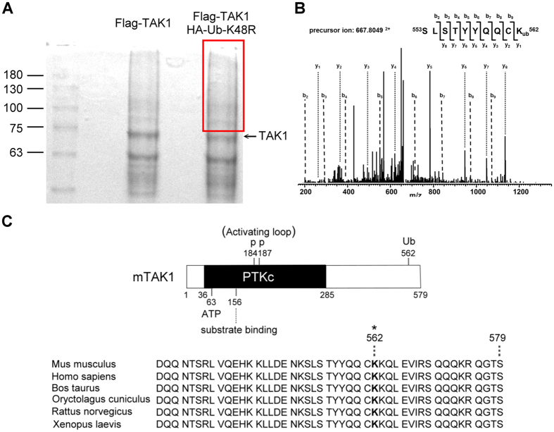
Figure 3. Lys562 residue of TAK1 is an ubiquitination site.
A, Coomassie blue stained gel indicates the band with TAK1 polyubiquitination. Flag-tagged TAK1 with or without HA-tagged ubiquitin were expressed in HEK293T cells. Cells were lysed, immunopricipitated, gel-separated, and stained with Coomassie blue. The square area indicated the presence of ubiquinated-TAK1. B, Lys562 residue of TAK1 was identified as an ubiquitination site by mass spectrometry. The protein sample with ubiquitinated TAK1 was recovered from the gel, enzyme digested, and subjected to mass analysis. Representative MS/MS spectra of peptides demonstrated ubiquitination at Lys562 of TAK1. Peaks matching expected b and y ions are labeled. C, Upper panel: schematic structure of TAK1, indicating functional domains and the new site identified in this study. Lower panel: alignment of part of the C-terminal of TAK1. The Lys562 residue is indicated by an asterisk.

