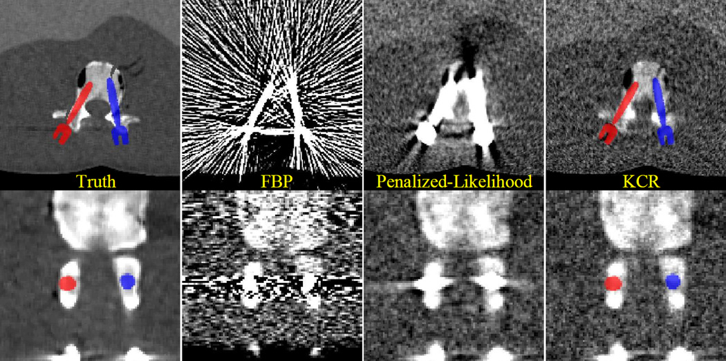
Figure 5.
A comparison of reconstruction approaches for the bilateral pedicle screw placement scenario. Severe streak artifacts are present in the FBP volume due to photon starvation in measurements containing rays passing through the pedicle screws. While the image is greatly improved using a statistical approach, artifacts remain in proximity to the devices, dramatically reducing the utility of the images for detecting breaches. In comparison, the KCR images are essentially streak-free and allow for visualization of anatomy very close to the implant. Specifically, the anterior simulated fracture is evident only in the KCR image. The lateral fracture remains difficult to visualize suggesting increased exposure/reduced regularization may be necessary.

