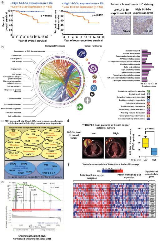
Figure 1. The clinical relevance of 14-3-3σ in breast cancer and tumour metabolic reprogramming.
(a) High 14-3-3σ expression was associated with prolonged breast cancer patients’ survival. 14-3-3σ protein expression levels in breast cancer patients at MD Anderson Cancer Center were evaluated by immunohistochemistry staining and matched with clinical data. 14-3-3σ protein expression in normal breast tissues was used as a reference to stratify patients.
(b) Down-regulation of 14-3-3σ is associated with upregulation of major cancer hallmarks especially deregulation of cellular energetics (i.e., metabolic reprogramming). Transcriptomic profiles of breast cancer patients (cohort GSE20194, n=255, Gene Expression Omnibus database) were analysed by Nexus Expression 3.0 software (BioDiscovery). The gene expression profiles of the lowest 14-3-3σ quartile were compared to those of the highest 14-3-3σ quartile and matched with corresponding biological processes and cancer hallmarks. A Circos map (circos.ca) was used to display the biological processes and cancer hallmarks that were upregulated upon 14-3-3σ down-regulation. The size of cancer hallmarks’ symbols indicated the magnitude of their upregulation upon 14-3-3σ silencing. The bar graph on the upper right corner shows a significant increase in cancer energy metabolism when 14-3-3σ expression is down-regulated. Enrichment scores were calculated based on transcriptomic analysis using Nexus Expression 3.0 program (BioDiscovery). Details about transcriptomic changes associated with 14-3-3σ down-regulation are described in Supplementary Figure X.
(c) Low 14-3-3σ expression is associated with increased oncogene expression and reduced tumour suppressors’ levels. This Venn diagram highlights genes and biological processes with significantly changed expression when 14-3-3σ is down-regulated in breast cancer patients’ tumours (cohort GSE20194, n= 255, Gene Expression Omnibus database).
(d) High 14-3-3σ expression is accompanied by a decrease in 18FDG uptake by human breast tumours. PET scan data and gene expression profiles of patients’ breast tumours (collected at MDACC) are combined in this analysis to study the impact of 14-3-3σ on tumour glucose uptake in patients. Average±95% confidence interval (CI).
(e–f) Gene Set Enrichment Analysis (GSEA, Broad Institute) of breast cancer patients showed upregulation of metabolic genes in breast tumours that expressed a low level of 14-3-3σ compared to breast tumours with high 14-3-3σ expression.

