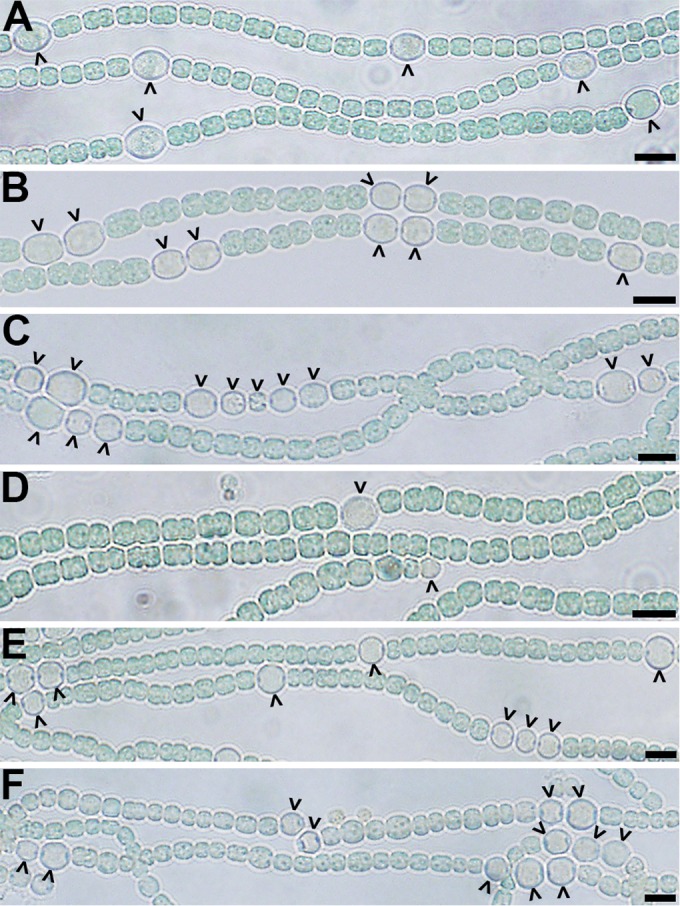
FIG 1.

Phenotypes of mutant strains. Representative bright-field micrographs of the wild-type (A), ΔpatS (B), ΔhetN (C), ΔhetC (D), ΔhetC ΔpatS (E), and ΔhetC ΔhetN (F) strains at 24 h (A, B, and E) or 72 h (C, D, and F) after the removal of combined nitrogen. Carets indicate heterocysts. Bar, 10 μm.
