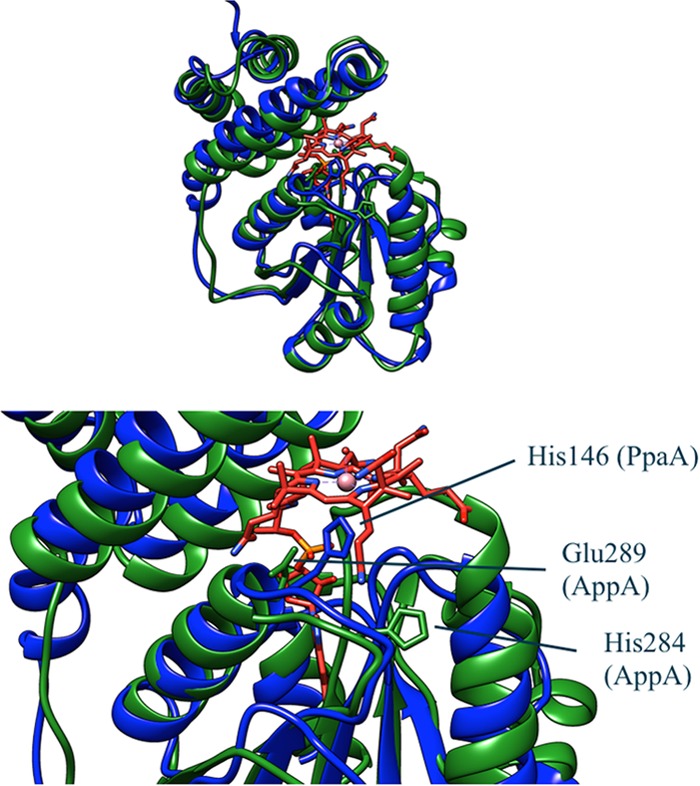
FIG 3.

Comparison of PpaA with the structure of the AppA SCHIC domain (Protein Data Bank accession no. 4HEH). PpaA is blue, AppA is green, and cobalamin is red. The structure of PpaA was predicted by the Phyre homology modeler. The strongly conserved histidine is shown in stick representation in both structures. The glutamate that replaces a strongly conserved glycine in AppA is also shown in stick representation. This glutamate is in close proximity to the phosphate group of the DMBI tail of cobalamin and may explain why AppA binds heme instead of cobalamin.
