
FIG. 5.
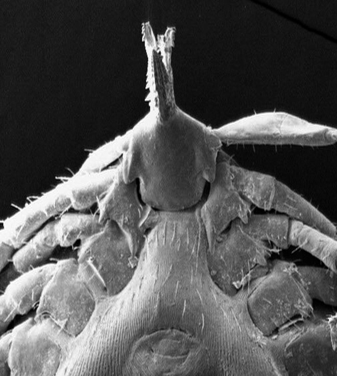
SEM of an Ixodes acuminatus tick collected from a rodent. Note the long, thin, straight inner spur of coxa I extending beyond anterior margin of coxa II, the triangular auriculae, and the fusiform hypostoma with thinly pointed tips.
Official websites use .gov
A
.gov website belongs to an official
government organization in the United States.
Secure .gov websites use HTTPS
A lock (
) or https:// means you've safely
connected to the .gov website. Share sensitive
information only on official, secure websites.
SEM of an Ixodes acuminatus tick collected from a rodent. Note the long, thin, straight inner spur of coxa I extending beyond anterior margin of coxa II, the triangular auriculae, and the fusiform hypostoma with thinly pointed tips.