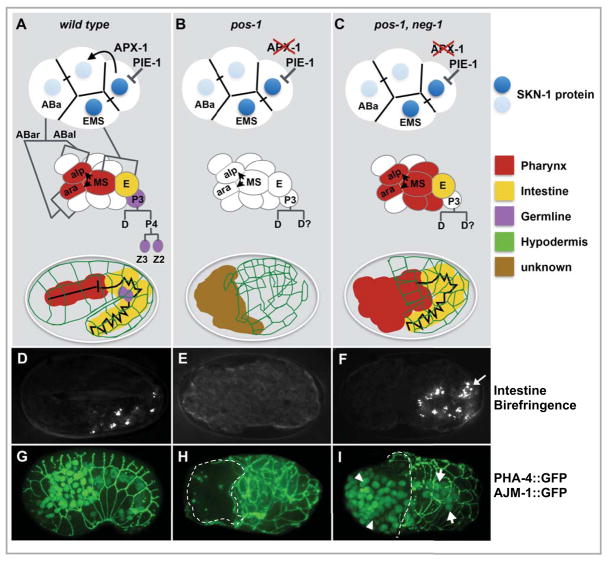
Figure 1. The pos-1 gutless phenotype is suppressed by neg-1 loss of function.
(A–C) Schematic diagrams showing key features in endo-mesoderm differentiation in wild-type and mutant backgrounds (as indicated). At the 4-cell stage SKN-1 localization and APX-1 signaling restrict endo-mesoderm potential to the blastomeres EMS and ABa, at the 12-cell stage MS signaling induces pharyngeal development in adjacent ABa descendants, finally body morphogenesis leads to enclosure of internal organs inside a network of hypodermal cells (diagrammed in green). The germ lineage P3 continues to divide asymmetrically producing the germline precursors Z2 and Z3 (purple). Defects in these events are indicated in the mutant contexts (B, C). (D–F) Polarized light micrographs showing gut differentiation as indicated by birefringent gut granule accumulation (D and F). (G–I) GFP fluorescence micrographs showing two differentiation markers: AJM-1::GFP which is expressed at cell-cell junctions of epidermal cells surrounding the embryo (McMahon et al., 2001), and PHA-4::GFP which is expressed in the nuclei of pharyngeal and intestinal precursors (Horner et al., 1998). In (H, I) failure of the AJM::GFP network to enclose the anterior region of the embryo is indicated by a dashed line, and PHA-4::GFP nuclear staining, absent in (H), is restored in pos-1, neg-1 double mutants (I, arrows).

