
Fig. 4.
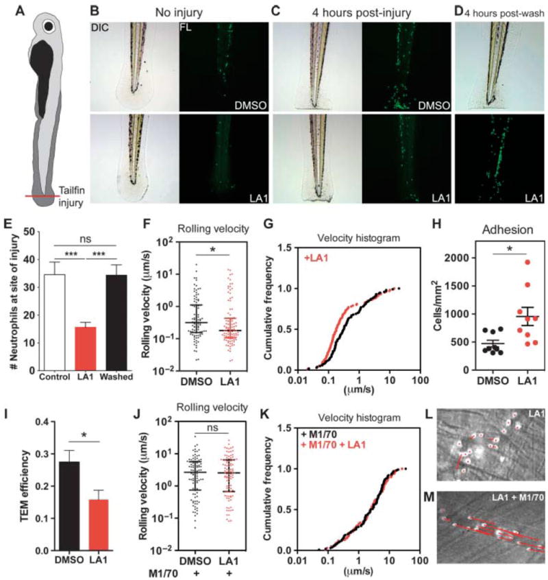
Leukadherins decrease leukocyte recruitment in vivo by increasing the extent of slow rolling and the number of adherent cells, thereby decreasing leukocyte TEM. (A) Schematic representing the zebrafish tailfin injury model. (B to D) Photomicrographs (left) and fluorescence images (right) of (B) the larvae tail without injury, (C) the tail with injury, and (D) 4 hours after the removal of LA1 from treated, injured larvae. Images show accumulation of neutrophils (green) in the injured tail. (E) Graph showing quantification of the number of neutrophils near the site of tailfin injury in zebrafish larvae treated with DMSO (Control) and LA1 (n = 12 to 16 larvae per group). Data are means ± SEM. ***P < 0.0001; ns, not significant (by one-way ANOVA). (F to M) Intravital microscopy–based determination of the effects of LA1 on leukocyte migration in vivo. (F) Graph showing the rolling velocities of individual neutrophils in the venules of TNF-α–treated mouse cremaster muscle without (DMSO) or with LA1. Lines indicate medians and 25 to 75% interquartile ranges. *P < 0.05. (G) Cumulative histograms of the rolling velocities of 100 leukocytes from DMSO-treated (black dots) and LA1-treated (red dots) animals. (H) Numbers of adherent neutrophils in venules without (DMSO) or with LA1. Data are means ± SEM. *P < 0.05. (I) Graph showing relative efficiency of neutrophil TEM (as determined by the number of transmigrated neutrophils/the number of adherent neutrophils) in the absence (DMSO) or presence of LA1. Data are means ± SEM. ***P < 0.05. (J) Rolling velocities of neutrophils in TNF-α– and M1/70-treated venules without (DMSO) or with LA1. Lines indicate median and interquartile ranges. ns, not significant. (K) Cumulative histograms of rolling velocity of 100 leukocytes from M1/70-treated mice in the absence (black dots) or presence of LA1 (red dots). Representative video micrograph images of cremaster muscle venules treated with (L) LA1 or (M) M1/70 and LA1. The lengths of the red arrows indicate neutrophil movement during a 25-s period.