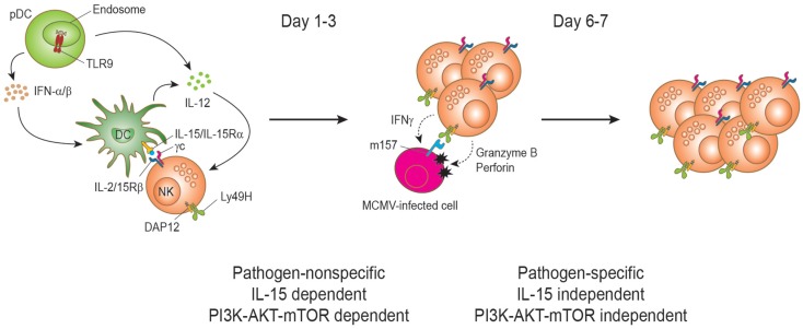
Figure 2.
Natural killer cell proliferation during murine cytomegalovirus infection. During MCMV infection, two stages of NK cell proliferation showing pathogen non-specific and specific responses have been proposed. IL-15 drives NK cell proliferation during the early phase of MCMV infection. Upon recognition of CpG motifs from MCMV viral DNA by TLR9, plasmacytoid dendritic cells (pDCs) secrete type I interferons (IFNα/β) and interleukin-12 (IL-12) cytokines. DC-derived IL-12 stimulates NK cells to produce IFN-γ. IFNα/β is transiently produced and reaches a peak level at day 1.5 post-infection and the production is important to induce the expression of IL-15. IL-15 is trans-presented by DCs to NK cells to induce proliferation and enhanced effector functions of NK cells. Activated NK cells can induce perforin/granzyme-mediated apoptosis of MCMV-infected cells by recognizing the viral m157 protein on the cell surface. This direct recognition depends on the activating receptor Ly49H. Proliferation at this stage is dependent on the PI3K–AKT–mTOR pathway. The interaction between Ly49H and the MCMV-encoded protein m157 drives the proliferation and expansion of Ly49H+ NK cells on days 6–7 post infection. Notably, this proliferation can occur in IL-15- and IL-15Rα-deficient mice during MCMV infection and is independent of the PI3K–AKT–mTOR pathway.

