. 2015 Jul 21;4:e07918. doi: 10.7554/eLife.07918

Figure 2. Telomerase activity is restricted by levels of TERT in differentiated cells while TR is limiting in wild-type hESCs.

(A) Targeting schematic of GFP, 3XFLAG-TERT (F-TERT), TR, and F-TERT+TR overexpression from the AAVS1 locus in wild-type hESCs. (B) Northern blot detection of total TR and 7SL in targeted hESC lines. TR runs as a doublet in UREA PAGE. (C) SDS-PAGE immunoblot of total TERT and tubulin proteins in editing hESC lines from whole cell extract. (D) TRAP assay of whole cell extracts from NPCs and fibroblast-like cells differentiated from GFP (G), F-TERT (T), TR (R), or F-TERT+TR (T&R) overexpressing hESCs using 200 ng protein. (E) Telomere restriction fragment assay of GFP, F-TERT, TR, and F-TERT+TR overexpressing hESCs.

DOI: http://dx.doi.org/10.7554/eLife.07918.007

Figure 2.

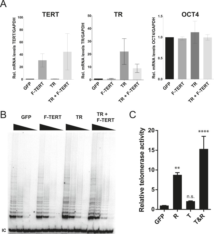
Figure 2—figure supplement 1. Quantification of TERT and TR expression levels and telomerase activity in the overexpression hESCs.

Figure 2—figure supplement 1.

(A) Quantitative RT-PCR of TERT, TR, and OCT4 in GFP, F-TERT, TR, or F-TERT+TR overexpressing hESCs. Expression level is relative to GFP hESCs and normalized to GAPDH. Error bars represent the SEM of three biological replicates taken 1 week apart. (B) TRAP assay of whole cell extracts from GFP, F-TERT, TR, or F-TERT+TR overexpressing hESCs using decreasing amount of protein (250, 50, 10, 2 ng). (C) Relative telomerase activity of the hESC lines was assayed by QTRAP. Values were set as fold activity relative to the GFP control line (n = 3, ANOVA with Tukey's test, SEM bars). R: TR, T: F-TERT and T&R: F-TERT+TR.