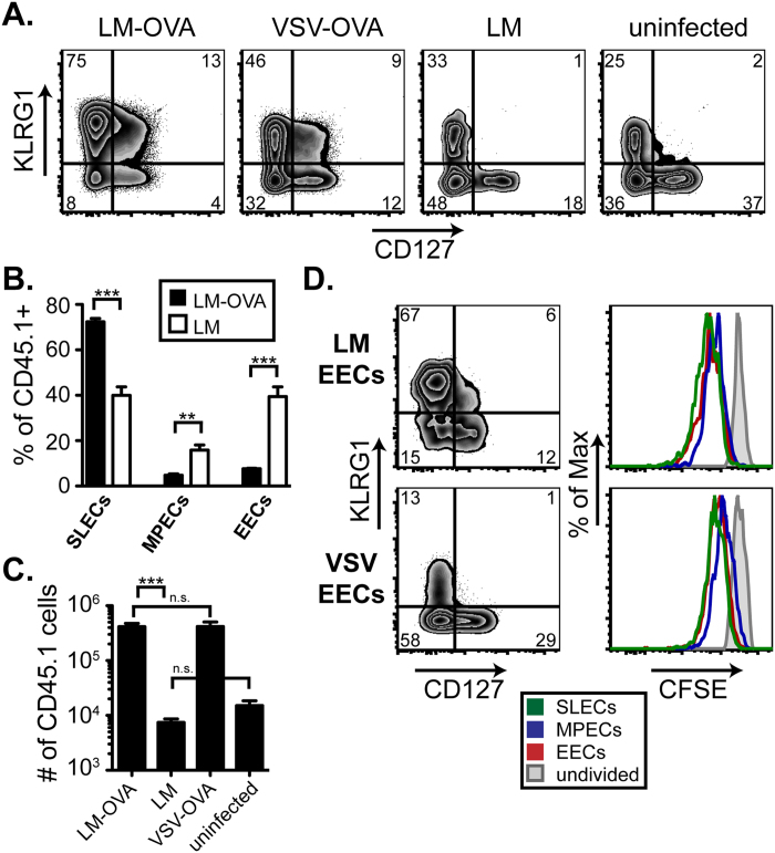
Figure 5. Optimal SLEC generation and proliferation requires continued antigen expression.
A. EECs were purified, as described in Fig. 2A, following VSV-OVA infection and transferred to recipient mice infected with LM-OVA, VSV-OVA, LM or left uninfected. 6 days following transfer, the CD8+, CD45.1+ EECs in the spleen were analyzed for KLRG1 and CD127 expression. Each condition was repeated at least 2 times. B. Percentages of SLECs, MPECs, and EECs are graphed for both LM-OVA EECs and LM EECs from 3–4 mice. C. Total numbers of CD8+ CD45.1+ transferred EECs per spleen, from the experiment in part A are shown graphically. D. EECs from both VSV-OVA- and LM-OVA-infected mice were sort purified as in Fig. 2A. Following purification, CD45.1+ EECs were mixed with CD45.2+ splenocytes from a naïve mouse at a 1:10 ratio prior to CFSE labeling. Total CFSE labeled cells were then transferred to uninfected recipients and CD8 CD45.1+ EECs were analyzed for KLRG1 and CD127 expression and CFSE dilution 3 days following transfer. The CFSE labeled CD45.2+ splenocytes were used as the undivided control.

