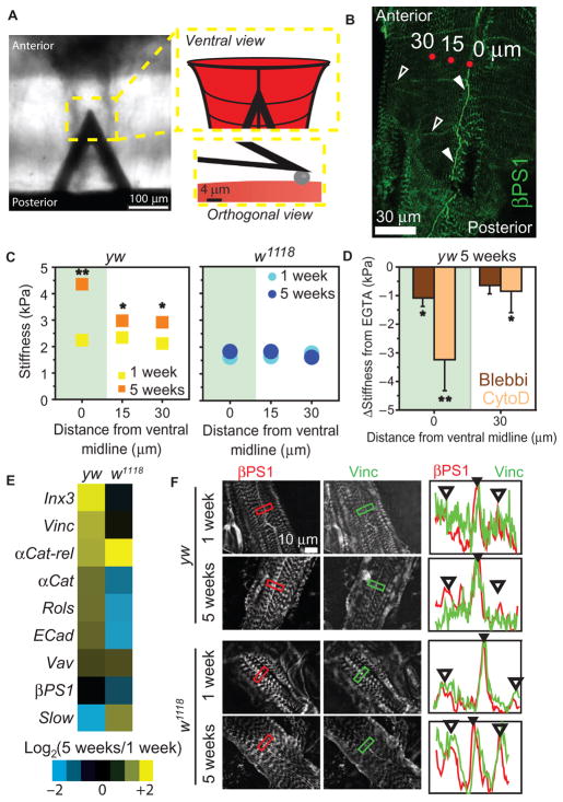
Fig. 4. Age-associated heart stiffening correlates with cortical actin cytoskeletal remodeling in flies.
(A) AFM cantilever positioned above the Drosophila heart. Insets are illustrations of a cantilever over the heart (top) and spherical indenter (bottom). (B) βPS1 localizes within hearts at IDs (filled arrowheads) and lateral junctions (open arrowheads) along the length of the fly heart. AFM indentation locations are indicated by red dots and their distance from ventral midline (0, 15, or 30 μm). (C) Cardiac stiffness as a function of distance from ventral mid-line in yw and w1118 flies (average ± SEM, n > 20). *P < 0.05, **P < 0.01 for 5-week versus 1-week at respective distance, using non-parametric t tests. Green overlay indicates ventral midline data. (D) Change in cardiac stiffness for 5-week yw after indicated pharmacological treatment. Data are individual flies with mean change in stiffness from EGTA ± SD (n = 10 per treatment). *P < 0.05, **P < 0.01, versus mean stiffness in EGTA using a repeated-measures one-way analysis of variance (ANOVA). (E) Ratio of aged (5-week) to adult (1-week) gene expression in the heart (n = 3 biological replicates of 10 pooled hearts per age and genotype). (F) Immunohistochemistry of hearts from 1- and 5-week flies showing vinculin or β1 integrin expression. Plots indicate fluorescence intensity from a line drawn within the box in each image for vinculin (green) or β1 integrin (red). Filled arrowheads indicate ID; open arrowheads, costameres.

