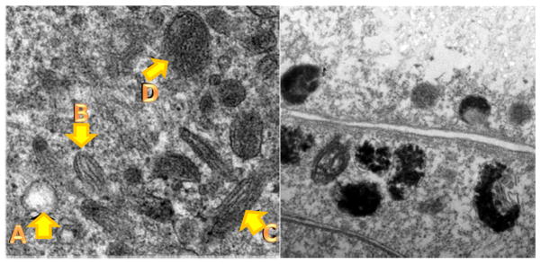
Figure 1. In situ transmission electron microscopy images of melanocyte and melanoma cells in culture.

Left panel, Stages of normal melanosomes in heavily pigmented melanocyte A: Stage 1, B: Stage 2, C: Stage 3, D: Stage 4. Right panel, abnormal melanosomes in MNT1 melanoma cells: note disruption of structure and difficulty in identifying stages. Adapted from our work Gidanian et al., 2008 [56].
