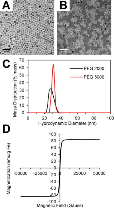
Fig. 1.

Characterization of iron oxide nanoparticles. (A) TEM image of SPIO cores and (B) PEG 2000 SPIOs negatively stained with phosphotungstic acid. (C) Hydrodynamic size distribution of SPIOs coated with DSPE-PEG 2000 and 5000 measured by dynamic light scattering. (D) Room temperature magnetization curve of SPIO cores.
