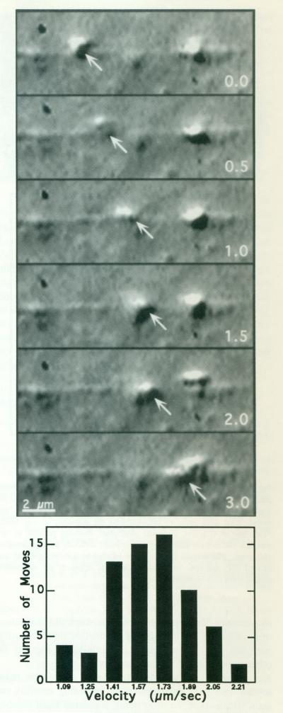
Fig. 2.
KI-washed organelles move on actin filaments. Upper: High-resolution DIC video sequence of a KI-washed organelle moving along an actin plume. This organelle moves, slows down at 1.5 s, and then speeds up again. Overall, it moves approximately 3 μm in 3 s. The actin plume was nucleated off the end of a KCl-washed acrosomal process, KI-washed, isolated organelles in 1/2× buffer were combined with pre-formed actin filaments in polymerization buffer containing ATP. The numbers in the right lower corner of each image represent the time in seconds. Lower: Movements of seven individual organelles moving on two different extended actin plumes were analyzed by measuring the distanced traveled during a ten-frame sequence in order to determine an instantaneous as opposed to an overall velocity. The ten frames were selected for inclusion if the organelle moved continuously from the first through the tenth frame; 69 such sequences were measured. Velocities were divided into categories according to the smallest incremental distance that could be measured on the video screen.

