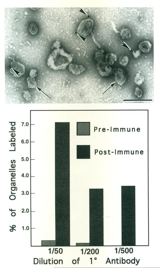
Fig. 3.
Colloidal gold labeling of KI-washed isolated organelles with anti-myosin antibody. Top: Micrograph of a representative field of negatively stained organelles defined by their limiting membrane (arrows). Four gold particles (arrowheads) are apparent in this field. Scale bar = 0.5 μm. Bottom: Histogram showing percent of organelles which were labeled by anti-myosin antibody or non-immune antiserum. The percent of organelles labeled by anti-myosin did not exceed 7%. Whereas very little label was apparent on organelles treated with non-immune antiserum.

