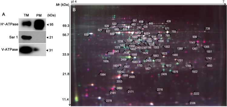
Fig. 3.
Purity assessment and a representative 2D-DIGE image. (A) Western blot analysis of extracted PM proteins with antibodies against proteins from different cell compartments. (B) PM proteins were separated by 2D-DIGE and detected with a Typhoon 9400 Scanner. The differentially accumulated protein spots under different NaCl and nitrate concentrations were determined using two-way ANOVA, and those positively identified by MALDI-TOF-TOF are marked by arrows.

