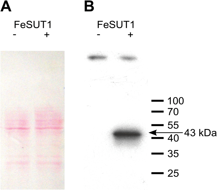
Fig. 2.
Western blot analysis of anti-FeSUT1 antiserum on isolated crude membrane protein extracts from yeast cells expressing FeSUT1 in sense (lane +) and in antisense direction (lane −). (A) Successful blotting was verified by Ponceau S staining. (B) The anti-FeSUT1-antiserum showed a strong specific band at 43kDa on membrane protein extracts from transgenic yeast that expressed FeSUT1 (lane +). The negative control showed no reactions (lane −). (This figure is available in colour at JXB online.)

