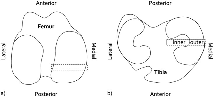
Fig 2. Osteochondral specimens to evaluate cartilage histopathological condition.

a) Location of the femur specimens. b) Location of the tibia specimens, displaying the in the inner and outer regions that were separately scored.

a) Location of the femur specimens. b) Location of the tibia specimens, displaying the in the inner and outer regions that were separately scored.