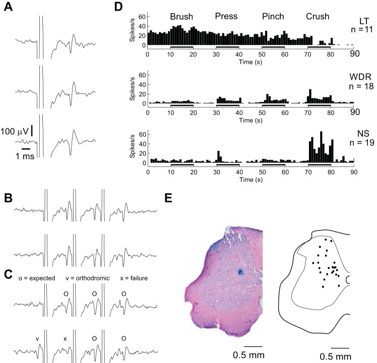
Fig. 3.
Characterization of recorded neurons. A–C: all included neurons met 3 criteria required for antidromic identification (Lipski 1981): neurons followed single pulse stimulation of the contralateral ventral cervical spinal cord (A), neurons followed trains of 3 pulses of stimulation at 200–333 Hz (B), and neurons exhibited orthodromic-antidromic collisions during activation of the neuron's peripheral receptive field (C). Axes in B and C are the same as in A. D: neurons fell into established low-threshold (LT), wide dynamic range (WDR), and nociceptive-specific (NS) categories as determined based on firing rates during brush-press-pinch-crush (BPPC) stimulation of peripheral receptive fields. E: neuron locations (right) based on 22 recovered Prussian Blue lesions (example left).

