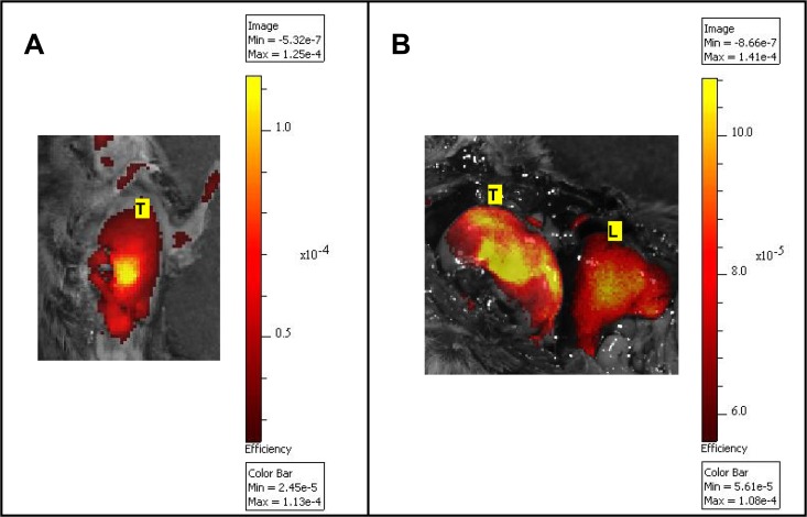
Fig 1. Imaging of MMP activity in the K-rasLSL-G12D lung cancer model using MMPSense680.
In vivo (A) and ex vivo (B) epi-fluorescent images (IVIS Spectrum) of a large superficial right lung tumor exposed after thoracotomy and laparotomy (B). The fluorescent bioactivatable probe reporting the proteolytic activity of MMP 2, -3, -9 and -13 (MMPSense680, 2 nmol) was injected i.v 24 h prior to imaging. The results confirmed the ability of an epi-illumination fluorescence system to image MMP activity in lung tumors, although the system could not discriminate large lung tumors from the liver (1A). Fluorescence intensity is represented as a pseudocolor image (MATLAB “hot” color map) overlaid on a white-light photographic image. Values of absolute fluorescence efficiency, as measured by the IVIS Spectrum, are shown on the color bar in units of 10−5 (1A) or 10−4 (1B). L: Liver; T: Right lung tumor.

