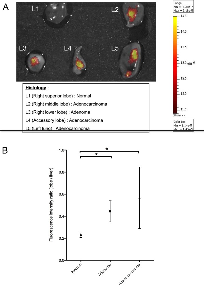
Fig 2. The fluorescent signal from MMPs increases in lung tumors.
A) Ex vivo image of MMP activity in superficial lung lesions of various stages from K-rasLSL-G12D mice. The nonneoplastic lobe (L1) of freshly isolated lungs imaged immediately after euthanasia did not display any fluorescent signal, while in the tumor-bearing lobes (L2-5), signal increased with the degree of lesion severity. The data confirmed the ability of an epi-illumination fluorescence system (IVIS Spectrum) to image MMP activity at different stages of lung tumor progression. B) Fluorescence intensity of lung adenomas (n = 8), adenocarcinomas (n = 3) and normal lung lobes (n = 25). In this experiment, the lung and tumor fluorescence signal was normalized on liver fluorescence, based on the hepatic activation of the probe. The fluorescence intensity ratio was calculated as a ratio of the median radiant efficiency of the lung lobe / liver. The median values ± interquartile range are shown, and the median differences between cancerous lesions and normal lobes are significant. * p value <0.05 by the Mann-Whitney test.

