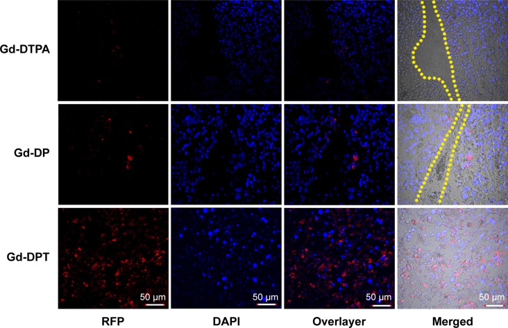
Figure 6.
Confocal luminescence microscopy showing the in vivo distribution and amount of RFP expressed after transfection by naked plasmid or nanoparticles. Upper panel shows Gd-DTPA/pRFP (Gd-DTPA), middle panel shows Gd-DP/pRFP (Gd-DP), and lower panel shows Gd-DPT/pRFP (Gd-DPT). Red indicates RFP, blue indicates DAPI-stained cell nuclei, and yellow lines indicate crevices in tumor tissue. Original magnification 200×.
Abbreviations: Gd, gadolinium; DAPI, 4,6-diamidino-2-phenylindole; pRFP, plasmid red fluorescence protein; DGL, dendrigraft poly-L-lysine; PEG, polyethylene glycol; DTPA, gadopentetate dimeglumine; DP, DTPA-DGL-PEG; DPT, DTPA-DGL-PEG-TAT.

