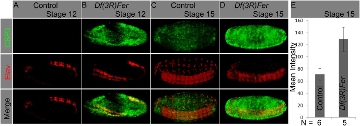
Fig 5. Ferritin mutants cause apoptosis in the CNS and other tissues.

(A-D) Whole embryos were stained with an α-CSP3act marking apoptotic cells (green), and an α-Elav marking neurons (red). Ectopic apoptosis was observed in ferritin mutants from stage 12 onwards; at this stage it was mostly restricted to the neurogenic region (B). At stage 15 apoptosis covers most mutant embryonic tissues (D). Quantification of the mean intensity value on CSP3act staining in control and ferritin mutants at stage 15 show a significant difference, with higher levels of staining in mutant embryos (n = 5; T-test, p = 0.0113).
