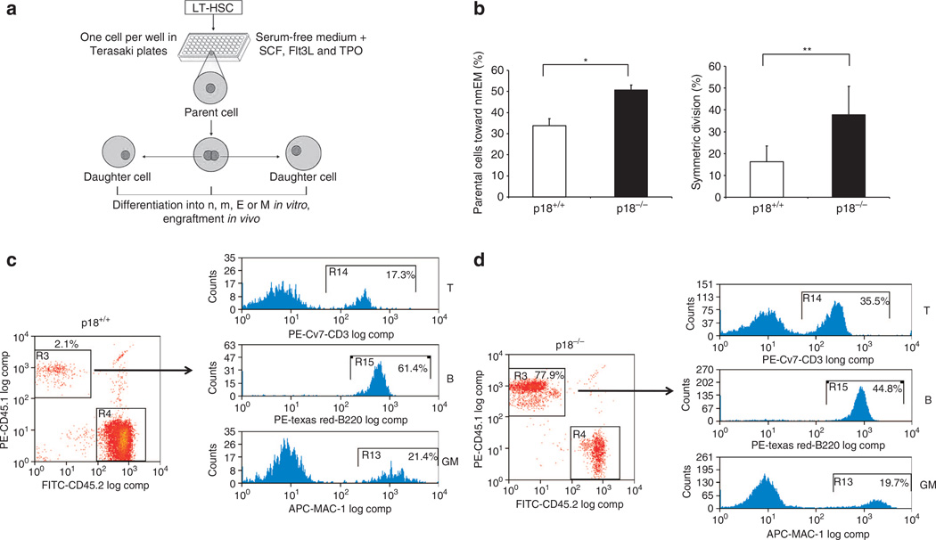
Figure 2. Increased probability of symmetric self-renewing division of HSC.
(a) Experimental approach of paired daughter stem cell analysis in vitro and multi-lineage engraftment of two daughter cells from a single HSC. (b) Percentage of parental HSCs committed to the nmEM lineage (left) and paired daughter HSCs able to form the nmEM progeny (right). (c) Representative recipient of the p18+/+ daughter cell. (d) Representative recipient of the p18−/− daughter cell. Multi-lineage differentiation was examined by flow cytometry. GM, T and B indicate lineages for myloid, T cells and B cells, respectively. Data sets were analysed using one-way analysis of variance (GraphPad Prism v6.0). All data represent mean ± s.d. in different groups. *P < 0.05, **P < 0.01.

