
Fig. 3. Initial expression of Hoxa13 does not occur in endothelial cells of the allantoic vasculature.
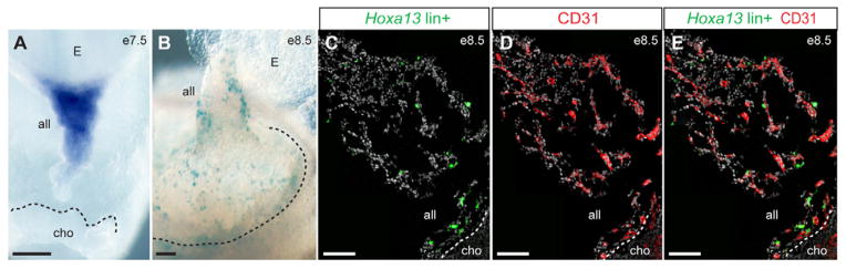
(A,B) Whole-mount X-gal staining of Hoxa13Cre/+;Rosa26R/+ mouse conceptus at E8.5 (B) reveals the fate of cells that have expressed Hoxa13 at E7.5 (A). Note the significant proportion of Hoxa13lin+ cells at the chorio-allantoic interface. (C–E) Immunostaining on allantois cryosections showing that most Hoxa13lin+ cells (green) do not express the endothelial cell marker CD31 (red) at E8.5. The mT/mG Cre reporter allele expresses GFP at the cell membrane and was used for colocalization with CD31, which is also expressed at the cell membrane. Nuclei are labeled with DAPI (gray). Dashed lines highlight the limit between the allantois (all) and chorionic plate (cho). E, embryo. Scale bars: 100 μm.