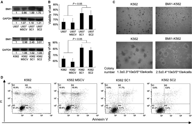
Figure 2.
BMI1 enhances the malignancy of leukaemic cells (A) Western blotting of BMI1 in subclones of transfected U937 and K562 cells. Numbers are the folds compared to the first line control. SC1 and SC2 represent two subclones of Bmi1 transfected U937 and K562 cells. (B) The viability of cell was examined by vi-cell XR cell viability analyser after cells were cultured without foetal bovine serum for 72 hrs, n = 3. (C) Colony forming assay of K562 and Bmi1-transfected K562. Cells were cultured in MethoCult®H4230 with 5*104cells/mL for 7 days and then replated two cycles. Numbers are colonies with diameter larger than 0.3 mm, n = 3. The colony number of Bmi1-transfected K562 was greater than control with statistical significance, P < 0.05. The colony number means the total number of colonies/number of cell input after two cycles replating. (D) The apoptosis ratio of K562 after 48 hrs of 5 μM arsenic trioxide treatment tested by annexin V and propidium iodide in flow cytometry.

