Figure 4.

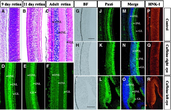
Pax6 expression pattern in the chick retina is perturbed by caffeine. Haematoxylin and eosin staining and Pax6 & HNK-1 immunostaining were performed on various developmental stage of chick retina. (A–F) Transverse sections of normal embryonic 9-day (A and D), 11-day (B and E) chick embryo and adult chick (C and F) retina stained with haematoxylin and eosin and Pax6 respectively. (G–R) Embryonic 9-day-old control (G–P) and caffeine-treated (H–R) retina stained with Pax6 and HNK-1 antibodies. Sections were counterstained with DAPI. The distribution of Pax6+ cells in caffeine-treated retina (N and O) is distinctly different from that of normal retina (M). INL, inner nuclear layer; ONL, outer nuclear layer and GL, ganglion cell layer. Scale bars = 500 μm in A–F, 100 μm in G and H, J and K, M and N, P and Q and 100 μm in I, L, O, R.
