Figure 2.

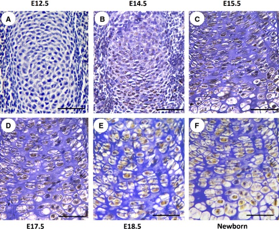
Immunohistochemistry of XBP1S in tibial growth plate chondrocytes in vivo. Temporal and spatial expression of XBP1S during chondrogenesis in tibial growth plates of post-coital day 12.5 mouse embryo (E12.5; A); post-coital day 14.5 mouse embryo (E14.5; B); post-coital day 15.5 mouse embryo (E15.5; C); post-coital day 17.5 mouse embryo (E17.5; D); post-coital day 18.5 mouse embryo (E18.5; E); and newborn (F) is shown. Microphotographs are shown of sections stained with anti-XBP1S antibody (brown) and counterstained with haematoxylin (blue). Immunostaining reveals positive nuclear staining in the entire chondrogenic developmental stages in both proliferating and hypertrophic zones. The scale bars represent 100 μm.
