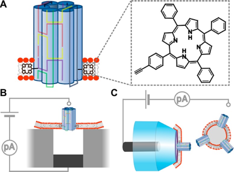
Figure 1.

DNA-based membrane-spanning pore and the two recording setups used to measure the conductance properties of the nanopore. (A) Schematic drawing of a six-duplex-bundle nanopore composed of six DNA oligonucleotides (colored) carrying two tetraphenyl porphyrin tags (black, inset), which anchor the pore into the lipid bilayer. The porphyrin anchor is attached via the acetylene group (inset) to position 5 of a uridine base (not shown). (B) Schematic drawing of a microcavity unit with a suspended planar lipid bilayer. Only one of the 16 units of the recording chip is shown. The nanopores were incorporated into the preformed lipid bilayers. (C) In the second recording setup, the nanopores were initially incorporated into lipid vesicles, which were then spread over the orifice of a 200 nm diameter glass nanopipette (transparent blue). The grounded reference electrodes in B and C are drawn as small, light gray circles, while the other electrode is in dark gray.
