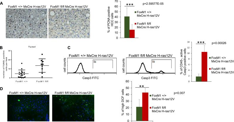
Fig. 3. Decreased proliferation, increased apoptosis and increased accumulation of ROS in tumors following deletion of FoxM1.
Tumor sections from mice (Alb-H-ras12V FoxM1+/+ MxCre and Alb-H-ras12V FoxM1fl/fl MxCre) following 5 injections of polyIpolyC were compared for PCNA expression by immunohistochemical staining (A). The tumor sections were also subjected to TUNEL assays (B); Single cell suspensions of the tumor tissues were compared also for active caspase3+ cells by flow-cytometry (C). Frozen sections of the tumor tissues were compared for accumulation of ROS following treatments with DCF-DA (D). The sections were treated with 10 μM 5-(6)-chloromethyl-2-dichlorodihydrofluorescein diacetate (CM-H2DCFDA) (Invitrogen, Carlsbad, CA) for 30 min at 37 °C and counterstained with DAPI. Images were taken using a fluorescent microscope at ×20 magnification.

