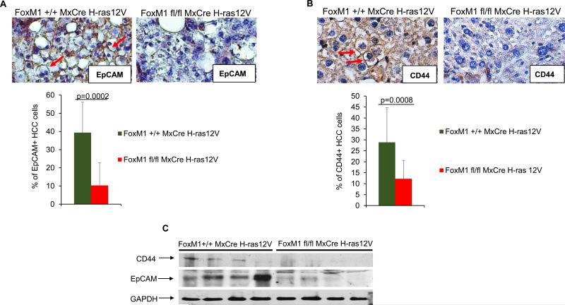
Fig. 4. Loss of the EpCAM+ and CD44+ HCC cells in the tumors following deletion of FoxM1.
Tumor sections from mice (Alb-H-ras12V FoxM1+/+ MxCre and Alb-H-ras12V FoxM1fl/fl MxCre) following 5 injections of polyIpolyC were compared for HCC cells that are positive for EpCAM (A) and CD44 (B). Quantification of the percentages of the EpCAM+ and CD44+ HCC cell over the total number of HCC cells are shown. For EpCAM+ cells, we quantified HCC cells from 9 different fields. For CD44+ cells, we quantified HCC cells from 16 different fields and 6 different tumors per genotype. (C) Protein extracts (150 ug) from tumor fragments were assayed by western blot.

