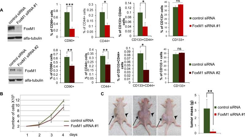
Fig. 6. Depletion of FoxM1 in Huh7 cells leads to specific loss of the cancer cells with stem cell features.
Huh7 cells were transfected with control siRNA, FoxM1-siRNA #1 and FoxM1 siRNA #2. (A) Western blots show the extent of FoxM1 depletion. The siRNA-transfected cells were treated with PE-tagged CD90-ab, FITC-tagged CD44-ab and PE-tagged CD133-ab and analyzed using a cell sorter. (B) Cell-growth following FoxM1-depletion was assayed by direct cell counting at the indicated times. (C) Twenty-four hours after control or FoxM1-siRNA transfection, 106 cells were subcutaneously injected into nude mice. The control-siRNA transfected cells were injected on the left side and the FoxM1-siRNA #1 transfected cells on the right side of 5 different mice. A picture of 3 mice after 6 weeks is shown. Quantification of the tumor mass from all five mice is plotted. Statistically significant changes were indicated with asterisks (*, p < 0.05; **, p < 0.01, ***, p<0.001.)

