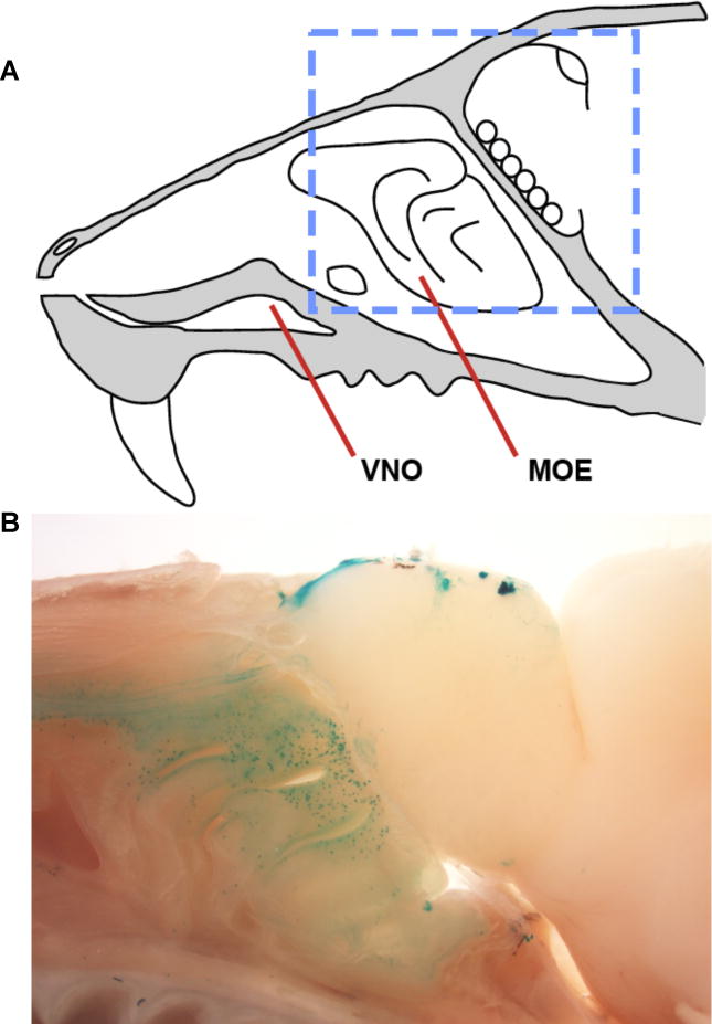
Figure 1. TAAR expression in the olfactory system.

(A) Anatomy of the mouse nose in sagittal view, with major chemosensory structures displayed (VNO- vomeronasal organ; MOE-main olfactory epithelium. (B) A sagittal section of olfactory tissue with TAAR-expressing neurons genetically labeled. Images are derived from TAAR5 knockout mice, in which β-galactosidase is expressed from the endogenous Taar5 locus. Labeled neurons include a subset of all TAAR neuron types (see discussion of receptor re-selection following Taar pseudogene expression), and are visualized by wholemount X-gal staining. The imaged region is indicated in panel A by the blue dashed box. Multiple TAAR-innervating glomeruli are observed in the olfactory bulb, as shown in Figure 4A. Image in panel A is adapted from [50].
