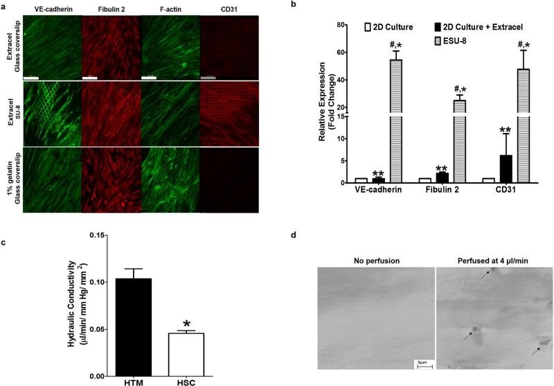
Figure 2. Characterization of HSC cell development on ESU-8 (12-μm pore) scaffolds.
(a) F-actin staining, revealing fiber alignment of the HSC cells on the Extracel-coated SU-8 scaffold. HSC cells cultured on the ESU-8 scaffold regained expression of cell characteristic marker, CD31, an expression that is lost in 2D culture on glass coverslips as well as exhibited proper expression and distribution of cell characteristic marker, VE-cadherin, when compared to 2D culture on glass coverslip. HSC cells cultured on the ESU-8 scaffold maintained expression of cell characteristic marker, fibulin 2. Scale bar = 100 μm. (b) qRT-PCR analysis confirming Extracel-induced upregulation of characteristic HSC markers on ESU-8 scaffolds. * (P< 0.001, ESU-8 vs 2D culture), # (P< 0.001, ESU-8 vs 2D culture + Extracel), ** (P<0.001, 2D culture + Extracel vs 2D culture). Mean ± SD with N=6. (c) Outflow study demonstrating hydraulic conductivity of the engineered SC inner wall was calculated to be 0.104 μl/ min/ mm Hg/ mm2, which is roughly 1/3 of the previously described biomimetic JCT layer of the HTM, an indication of endothelial integrity. (d) SEM images revealing formation of vacuole-like micron-sized pores in the engineered SC inner wall following perfusion at 4 μl/ min for 6 h.

