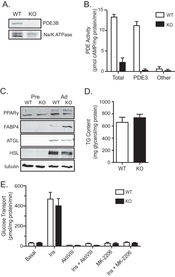
FIG 2.

Characterization of WT and Pde3b-KO brown adipocytes. (A) Immunoblot for PDE3B protein in crude membrane lysates harvested from WT and Pde3b-KO brown adipocytes. Na/K ATPase, a membrane-bound protein, was used as a loading control. (B) A radioactive assay for PDE activity was performed on membrane lysates. For the PDE3-specific activity, 10 μM cilostamide was added to the reaction mixture for both WT and Pde3b-KO adipocytes, and the activity was subtracted from total activity. Other cAMP-PDE activity was calculated based on inhibition after the addition of 100 μM IBMX. (C) The protein expression of peroxisome proliferator-activated receptor gamma (PPARγ), fatty acid binding protein 4 (FABP4), adipose triglyceride lipase (ATGL), and hormone-sensitive lipase (HSL) for the two genotypes was compared via Western blot analysis. (D and E) Triglyceride (TG) content (n = 3) (D) and glucose transport (n = 3) (E) were measured in WT and Pde3b-KO adipocytes. All data are presented as means ± standard errors of the means.
